

Chapter 5: Table of Contents
Cranial Abdominal Incision and Falciform Ligament
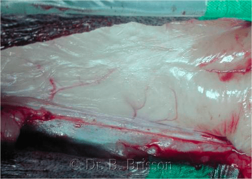
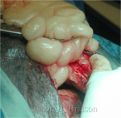
When using a cranial abdominal incision, the falciform ligament is removed to improve cranial abdominal visualization; this is especially important in overweight patients and when cranial abdominal procedures such as those involving the liver, stomach and spleen are being performed. Once the abdominal cavity is open, the falciform ligament lays like a blanket cranio-caudally between the xiphoid and the umbilicus. The falciform ligament is attached to the body wall cranially along the xiphoid and laterally on either side of the body wall but not caudally where it can be elevated at the level of the umbilicus to allow its removal.
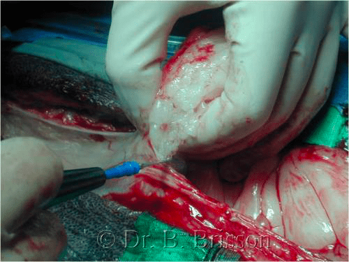
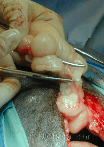
To remove the falciform ligament, transect it at its attachment on either side of the linea alba with scissors or cautery and ligate or cauterize any bleeding vessels. After releasing the lateral attachments, ligate the base of the falciform ligament, like a pedicle, close to the xiphoid with absorbable monofilament (size 2-0 or 0) using a circumferential or transfixing ligature. The fatty ligament can then be amputated just distal to the ligature. Simple avulsion or separation of the falciform ligament is possible but may limit visualization or lead to unnecessary hemorrhage. The falciform ligament is not encountered and does not need to be removed when a caudal abdominal incision is performed (e.g. cystotomy only).
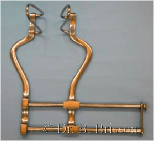
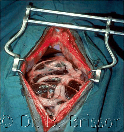
Once the falciform is removed, insert a Balfour abdominal retractor of appropriate size to maintain abdominal wall retraction – this is a very helpful instrument for surgeons working without an assistant!