

Chapter 21: Table of Contents
Intraluminal Tracheal Stenting
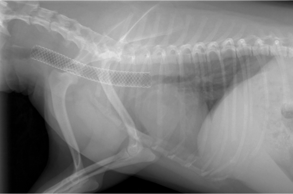
Intraluminal tracheal stenting is a newer, minimally invasive, interventional technique that consists of placing an inert metal stent within the trachea under fluoroscopic guidance using an oropharyngeal approach. During the last 15 years, several intraluminal stents have been used for this purpose with varied success. Balloon expandable biliary stents and stainless steel, self-expanding stents were used in initial reports. At present, the Vet Stent-Trachea® sold by Infiniti Medical is considered the stent of choice for this procedure. Vet Stents are self-expanding, flexible, woven stents made of nitinol. Nitinol is a nickel (NI) and titanium (TI) alloy. It is referred to as a shape memory metal since it can be given a shape when warmed, deformed when cooled (flattened and straightened to be placed into a delivery system) and it will return to its predetermined shape and size when deployed at body temperature. Newer veterinary stents include the Duality stent® which offers a dual diameter to accommodate large tracheal diameter discrepancies (cervical vs intra-thoracic).
Unlike extraluminal rings, intraluminal stenting allows treatment of both cervical and intrathoracic tracheal collapse without surgical incision. Because there is little long-term (>5 years) follow-up of patients who have undergone intraluminal stenting and because complications are relatively frequent and can be serious, tracheal stenting is currently indicated as an end-stage option for patients with intrathoracic tracheal collapse who have failed medical management or for patients who are not considered good candidates for surgery. It is unclear whether extraluminal rings or intraluminal stents are better for patients with cervical collapse alone. Mainstem bronchus collapse is not typically treated using intraluminal stents. It is important to note that being unable or unwilling to administer oral medications for life is not a good reason to recommend stenting since most patients who receive a stent still require medical therapy for life.
Intraluminal stents are inserted under general anesthesia using fluoroscopic guidance. Laryngeal examination should be performed at induction to identify laryngeal paralysis/collapse and elongated soft palate. If laryngeal paralysis is documented (rare), a laryngeal tie-back should be performed. A staphylectomy should be performed for an elongated soft palate. For stenting, the dog is positioned in lateral recumbency on the fluoroscopy table. A marker catheter is inserted in the esophagus to account for radiographic magnification. In lateral recumbency, and after retraction of the endotracheal tube to the level of the larynx, images are collected at rest, under positive (and if possible negative pressure) ventilation in order to determine the maximal tracheal diameter as well as the length and location of the collapse. A stent that is approximately 10-20 % larger than the tracheal diameter and of appropriate length (at least 1cm beyond at either end of the collapse but ideally spanning most of the trachea) is chosen and inserted into the trachea. Vet Stents are flexible, self-expanding, nitinol stents. They are reconstrainable, which means that if less than 75-80% of the stent has been deployed it can be recaptured within the delivery system to allow repositioning. Stents cannot be re-captured once fully deployed (they can however be removed in the days that follow stenting if absolutely required but cannot be reused). Stents should be placed at least 5mm caudal to the cricoid and cranial to the carina to prevent laryngospasm / excessive coughing and obstruction of the mainstem bronchi respectively. Anesthesia for these cases can be intense and requires good communication between the clinician performing the procedure and the anesthetist.