

Chapter 20: Table of Contents
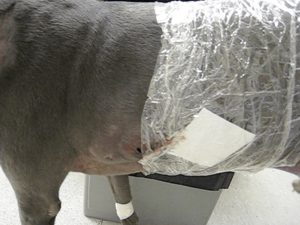
During initial assessment, protect the wound from further contamination by applying a sterile dressing (i.e. non-adhesive gauze sponges) and light soft padded bandage. If the injury appears to communicate with the thoracic cavity, seal the wound by covering it with a sheet of commercially available medical adhesive film and if unavailable any type of clean plastic film (e.g. plastic bag or kitchen plastic wrap). If hemorrhage is present, local pressure or vessel ligation may be necessary. Digital pressure or by application of a soft padded bandage is preferred. A tourniquet may be considered if direct pressure fails to control active arterial hemorrhage. An Esmarch tourniquet bandage may be used over the forefoot or hindfoot, and is applied by wrapping elastic or soft rubber bandage (e.g., Vetrap) tightly over the foot, from digits stopping just proximal to the wound. A window is then created in the bandage over the wound, exposing the tissues and allowing identification and ligation of vessels. The Esmarch bandage is then promptly removed. A tourniquet is used for the purpose of identifying and ligating vessels that continue to hemorrhage despite the application of direct pressure. Note: If any form of tourniquet is used to control hemorrhage of a distal extremity, it should be placed distal to the elbow or stifle joint and its use limited to <20 minutes at one time. Use caution and note that nerve damage or ischemic injury may result from tourniquet application.
Thoracic focused assessment with sonography for trauma (tFAST) and abdominal FAST (aFAST) examinations are useful to quickly assess for free thoracic or abdominal fluid. Once the patient is stabilized, thoracic radiographs can be performed. These are recommended for any wound or puncture over the neck, thorax or abdomen. Evaluate the pleural cavity for pneumothorax (if communicating thoracic wall wound or perforated lungs), pleural effusion (hemorrhage or pyothorax). Evaluate the lung parenchyma for atelectasis, consolidation or lung lobe torsion. Evaluate ribs for fractures and vertebral bodies for fracture/subluxation injuries. *The absence of pneumothorax does not preclude penetrating thoracic injury with communication to the pleural cavity.
Abdominal radiographs are performed to evaluate for free air in the peritoneal cavity (indicating perforated bowel or full-thickness abdominal wall wound) and lack of serosal detail in the retroperitoneal space (indicating uroabdomen, hemoabdomen, bile peritonitis, septic peritonitis). Identifying the urinary bladder does not rule-out urinary trauma but is a good sign. Organ herniation may also be identified on abdominal radiographs (e.g. abdominal wall hernia or pre-pubic tendon injury).
Radiographs of the extremities are recommended for wounds of the lower extremities since the underlying bones and ligaments may be injured (especially with shearing or bite wounds). It is important to perform radiographs of a region where there is suspicion of associated bone or ligament injury. Shearing injuries over the carpal or tarsal joint may also warrant stress radiographic views to detect ligamentous instability; however, this should only be performed once the animal is stable and has sufficient analgesia.