

Chapter 12: Table of Contents
Wedge Biopsy Technique
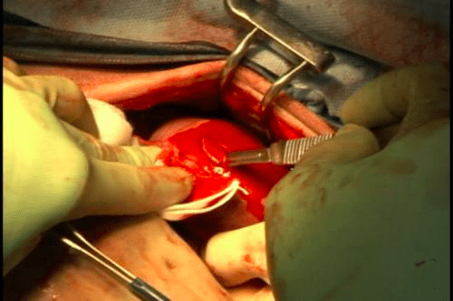
To obtain a wedge biopsy, an assistant should gently occlude the renal hilus by clamping it between an index and middle finger to reduce renal blood flow. The surgeon stabilizes the kidney with the non-dominant hand and incises the kidney with a scalpel held in the dominant hand. A wedge of tissue is removed (~1cm long, 0.5 cm wide). Significant hemorrhage should be expected. Quickly apply pressure to the biopsy site and prepare to appose the biopsy edges. Using small diameter, monofilament absorbable suture, place a simple interrupted or simple continuous suture pattern along the incised edges. Use a surgeon’s throw and slowly tighten the suture to appose the cut edges; rapid apposition often results in tearing due to the friable nature of the renal capsule and parenchyma. Apply pressure for a few more minutes if the bleeding persists.