


Chapter 6: Table of Contents
Suspensory Ligament
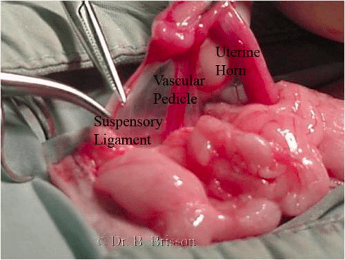
Once you have retrieved the left uterine horn, apply the tip of a curved mosquito forceps to the proper ligament (located between the ovary and the uterine horn). Grasp the ovary between your thumb and index finger (palming the mosquito forceps with the same hand) and apply caudal and ventral traction (towards the caudal abdomen and ceiling) to identify the suspensory ligament as a taut fibrous band located at the cranial aspect of the ovarian pedicle. To allow further exteriorization, stabilize the ovary and mosquito forceps with your right hand (if you are a right handed surgeon) and use your left index finger to palpate, stretch (caudomedial traction), and break the suspensory ligament. The orientation of the suspensory ligament is caudo-cranial from the ovary towards the kidney and that of the ovarian pedicle is ventrodorsal. For this reason, it is safest to break the suspensory ligament deep into the abdomen (away from the ovarian tissue and vascular pedicle). The left suspensory ligament can often be visualized while the right is often too short to be exteriorized (especially in deep chested dogs). If the ligament can be visualized, you can break it using mosquito forceps or cut it with Metzembaum scissors.